
Mediastinal & Hilar Lymphadenopathy
Expert Evaluation of Enlarged Chest Lymph Nodes
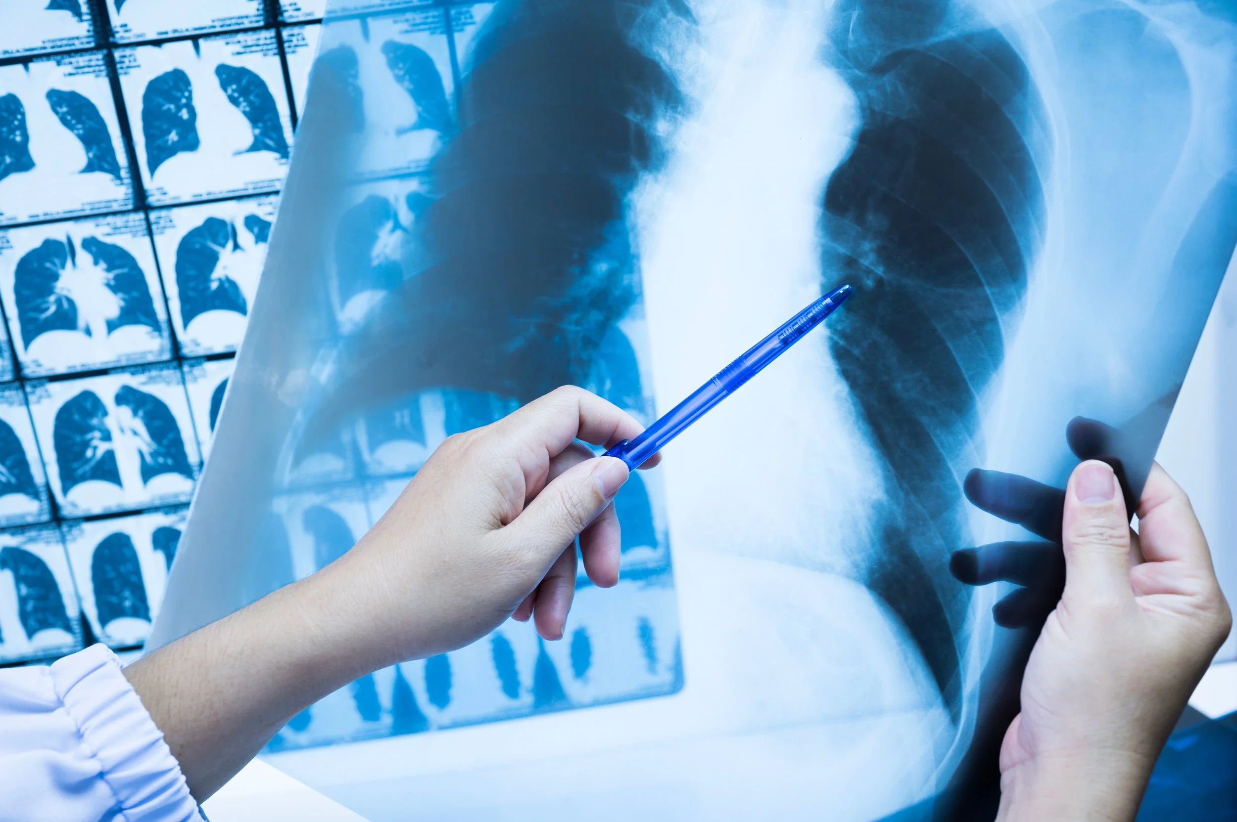
Mediastinal and hilar lymphadenopathy refers to enlarged lymph nodes located in the center of the chest (mediastinum) or around the lung roots (hilar region). These lymph nodes play an important role in immune defense, but when they become enlarged, it may signal infection, inflammation, autoimmune disease, or malignancy.
Enlarged chest lymph nodes are often discovered incidentally on imaging such as a chest X-ray or CT scan. In other cases, they are identified during evaluation of symptoms like persistent cough, shortness of breath, fevers, or unexplained weight loss.
Common Questions About Mediastinal & Hilar Lymphadenopathy
-
Lymphadenopathy means enlarged lymph nodes.
The mediastinum is the central compartment of the chest between the lungs, and the hilar regions are where the airways and blood vessels enter the lungs. When lymph nodes in these areas become enlarged, it may indicate that the immune system is responding to an infection, inflammatory condition, or abnormal cell growth.
Enlargement alone does not confirm a diagnosis — it signals the need for further evaluation.
-
There are several potential causes, ranging from mild to serious.
Common causes include:
Viral or bacterial infections
Fungal infections
Sarcoidosis
Tuberculosis
Autoimmune conditions
Lymphoma
Metastatic cancer
Primary lung cancer
The pattern, size, symmetry, and associated imaging findings help guide diagnostic suspicion, but tissue sampling is sometimes necessary for definitive diagnosis.
-
These lymph nodes are typically detected on imaging studies such as:
Chest X-ray
CT scan of the chest
PET scan
Often, they are discovered incidentally during imaging performed for another reason. In other cases, they are found during evaluation of symptoms such as chronic cough, abnormal lung imaging, or suspected malignancy.
-
No. Many cases of mediastinal or hilar lymphadenopathy are caused by infections or inflammatory conditions rather than cancer.
However, certain imaging characteristics — such as large size, asymmetric enlargement, PET scan activity, or associated lung nodules or masses — may increase concern and warrant further evaluation.
Because imaging alone cannot always determine the cause, additional testing is sometimes required.
-
Diagnosis begins with a detailed review of imaging, medical history, symptoms, and risk factors.
If further evaluation is needed, minimally invasive biopsy techniques are often recommended. The most common diagnostic procedure is:
Endobronchial Ultrasound (EBUS)
EBUS is a specialized bronchoscopy procedure that allows real-time ultrasound-guided biopsy of mediastinal and hilar lymph nodes through the airways.
It is performed under sedation and does not require surgical incisions. EBUS provides tissue samples that can help diagnose conditions such as sarcoidosis, lymphoma, infection, or cancer.
In select cases, additional testing such as blood work, PET imaging, or surgical biopsy may be necessary.
-
Treatment depends entirely on the underlying cause.
Management may include:
Monitoring with repeat imaging
Antibiotics or antifungal therapy
Treatment for sarcoidosis or autoimmune disease
Oncology referral and staging if malignancy is identified
Coordination with infectious disease or thoracic surgery when appropriate
Our role is to establish an accurate diagnosis and guide patients toward the most appropriate, evidence-based care pathway.
-
Referral is recommended when:
Enlarged mediastinal or hilar lymph nodes are identified on imaging
PET scan shows hypermetabolic lymph nodes
There is concern for sarcoidosis, lymphoma, or metastatic disease
Tissue diagnosis is required
Abnormal lymph nodes are associated with lung nodules or masses
Symptoms such as persistent cough, weight loss, fevers, or night sweats are present
Timely pulmonology evaluation allows for efficient diagnostic workup and can prevent unnecessary delays in treatment.
At The Lung Docs, we provide advanced diagnostic bronchoscopy, including EBUS-guided biopsy, to deliver accurate answers and coordinated specialty care.
Find a Location Near You
The Lung Docs provides specialized, state-of-the-art pulmonary care to our patients with asthma in Chattanooga and the surrounding Southeast Tennessee and Northwest Georgia areas.

PULMONOLOGIST
Dr. Mike Czarnecki
I’m Dr. Mike Czarnecki, “The Lung Doc,” and I’m trained in all areas of pulmonary health, including the diagnosis and treatment of mediastinal & hilar lymphadenopathy. I will work with you to formulate a personalized treatment plan so you can live, laugh, love, and breathe better again! To book an appointment with me, call 423‑710‑3864or request an appointment online. I can’t wait to meet you!